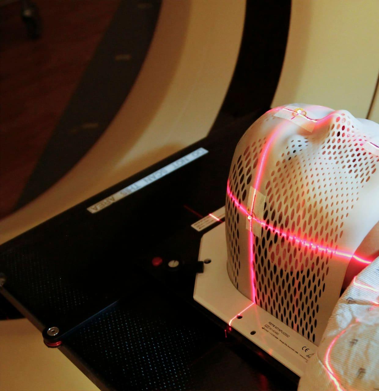
Limited labelled medical data
Expert annotation is slow and costly, and access to imaging is tightly governed.
Medical Imaging Research
Investigating image augmentation, segmentation, annotation consistency and model robustness in controlled medical imaging research workflows.
Research methodology only. Not a medical device, no clinical validation, and no Synthex results published yet.
Research problem
The questions this project investigates, stated as problems rather than solved claims.
Expert annotation is slow and costly, and access to imaging is tightly governed.
Small, faint or irregular lesions are easy to miss and hard to delineate.
Experts can disagree on whether, and where, a lesion is present.
Training and deployment data can differ in population, protocol and equipment.
Dose, reconstruction and slice thickness change how images look.
Good scores on one dataset may not transfer to another.
Research workflow
The intended experimental workflow. Each stage produces a recorded artefact so results can be reproduced and questioned.
STEP 01
Public, de-identified collections used under their licence terms (e.g. LIDC-IDRI, CC BY 3.0).
STEP 02
Documented resampling, intensity windowing and region-of-interest extraction.
STEP 03
Reference masks derived from expert annotations, with disagreement retained.
STEP 04
Controlled variations added to the training split only; never to evaluation data.
STEP 05
Fixed architecture, configuration and recorded random seeds per run.
STEP 06
Dice, IoU, precision, recall and sensitivity on held-out real images.
STEP 07
Failures grouped by lesion size, appearance and acquisition condition.
Evaluation stays real
Not a clinical pipeline
Imaging data explorer
Switch views and evaluation focus to see what a researcher inspects. Original schematic drawings and static text — no patient data, no collection images, no model output.
Illustrative Imaging Explorer — Schematic, Not Patient Data
Illustrative DemonstrationOverlap (agreement), false-positive area (predicted only) and false-negative area (missed).
What a researcher inspects
Metrics used (names only)
Static fixture: an original schematic drawing and descriptive text. No patient data, no images from any collection, and no model output. Not a clinical tool.
Experiment comparison
The comparison protocol for this project. Values appear here only once a result is verified and approved in the evidence register — none are yet.
Comparison protocol
Evidence: Unavailable · R-003What changes
Nothing — trained on real annotated images only.
Held fixed
Splits, architecture, preprocessing, training budget, seeds.
Question answered
What does the model achieve without synthetic data?
Results matrix
| Configuration | Dice | IoU | Precision | Recall | Sensitivity |
|---|---|---|---|---|---|
| Real-data baseline | No verified result | No verified result | No verified result | No verified result | No verified result |
| Real + synthetic augmentation | No verified result | No verified result | No verified result | No verified result | No verified result |
| Alternative augmentation | No verified result | No verified result | No verified result | No verified result | No verified result |
| Challenging evaluation conditions | No verified result | No verified result | No verified result | No verified result | No verified result |
No results are published: no experiment has been verified and approved in the evidence register. Values will appear here only when they are.
Annotation quality
Evaluation is only as reliable as its reference masks. This is the proposed audit workflow.
Visible lesions with no mask, which make correct predictions look like false positives.
Masks that differ at the edges, which matters most for small lesions.
Empty, fragmented or anatomically implausible shapes.
Measured agreement between readers, kept rather than averaged away.
Flagged cases ranked so expert time goes where it matters.
Re-checks after preprocessing and augmentation to catch drift.
Empty, fragmented and implausible masks; agreement between readers.
Cases scored by how likely the label is to be wrong.
Qualified reviewers accept, correct or reject each flagged label.
Outcome and rationale stored with the dataset version.
Expert review queue
Illustrative · static fixture| Case | Automated flag | Priority |
|---|---|---|
| case-A | Readers disagree on lesion presence | High |
| case-B | Mask fragmented into 3 regions | Medium |
| case-C | Boundary differs between readers | Medium |
| case-D | Mask empty; model detects a region | High |
Placeholder case labels only. A real queue would hold no patient-identifiable information on any public surface.
Research limitations
Synthetic images may differ from real scans in ways models exploit.
Reference masks reflect specific readers, protocols and conventions.
Public collections may not represent other populations, scanners or sites.
Results on one collection need confirmation on independent data.
Clinical experts must review labels, failure cases and interpretations.
Dice or sensitivity do not establish clinical benefit, safety or readiness.
References
Public sources this research direction refers to. Synthex is not affiliated with these organisations, and no images from these collections are reproduced on this site.
Public thoracic CT collection with radiologist lesion annotations. Licence: CC BY 3.0; TCIA Data Usage Policy applies.
The Lung Image Database Consortium (LIDC) and Image Database Resource Initiative (IDRI) — primary publication.
Data citation, The Cancer Imaging Archive.
Licence: LIDC-IDRI is distributed under CC BY 3.0 subject to the TCIA Data Usage Policy and Restrictions.
Research pilot
We welcome research collaborations on annotation quality, robustness evaluation and carefully baselined synthetic augmentation. Please do not send patient data through this website.
No medical device certification, clinical approval, NHS partnership or validated patient outcome is claimed.